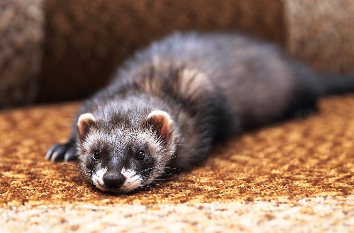
Like it or not, admit it or not, poop has fascinated us over the years. When poop is good, we hardly notice, but when it is bad, we pay attention.
About 10 years ago, I wrote a short article for the Internet called “The Poop Sheet” (now called “The Poop Chart”). I really stepped in it that day, and this particular subject seems to have followed me for years. People have reprinted the “Poop Chart” over and over, and I have even been asked to lecture on ferret poop.
Here is the full, unadulterated story of “the poop, and nothing but the poop.”
Basic Rules Of “Poop-ology”
Each segment of the gastrointestinal tract of ferrets (and all other animals) has a specific role in processing food and making its components available for use by the body. The first rule of “poop-ology” is one of focus: the end goal of the digestive process is nutrition, not the production of poop.
Another very important concept, and one that I often have to remind ferret owners of, is “Don’t be a stool-gazer.” This means, don’t ascribe too much importance to an individual poop. Most ferrets will poop three or four times a day. One bad one is not the end of the world, and one good one, in an animal with GI problems, doesn’t necessarily mean that all is forgiven.
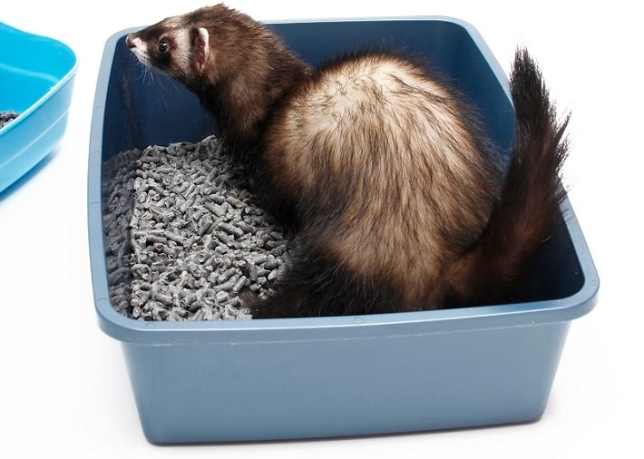
Why look at ferret poop, anyhow? Well, you are there cleaning the litter anyway, and it’s always nicer to perform a medical chore than just a janitorial one. Although the condition of the poop is not a very specific indicator of overall health, it can be an early warning sign for many forms of gastrointestinal disease. Often the bowel movements of ferrets herald systemic disease far earlier than any other signs.
Three components — color, shape and consistency — are used to characterize ferret poop and might help determine where a problem in the G.I. tract might exist.
Normal ferret poop is a light tan to brown color; has a smooth, toothpaste-like consistency and is tubular in shape. Exposure to air allows it to dessicate, thus causing it to shrink, turn dark brown and get hard.
It’s important to note that a lot of diseases, especially non-GI diseases, allow ferrets to pass totally normal feces, so good poop doesn’t equate totally with perfect health. Keep that in mind.
The Ride Begins
The gastrointestinal tract is a long tube in most animal species, with distinct sections in which various stages of digestion take place. Each section performs a specific function, and failure of any part often affects the poop in different ways. Some areas are associated with subtle changes, some with more obvious ones.
The first stop is the oral cavity. In this area, both the teeth and the saliva cause profound and important changes to the food. Teeth, of course, are important for grabbing and crushing the food. The type of diet determines how the food is processed.
Due to the hard-packed nature of kibble, part of essential ferret supplies, each piece must be broken into much smaller pieces so that digestive fluids such as saliva and gastric acids can begin the digestive process. Advanced periodontal disease may limit a ferret’s ability to crush and swallow its food properly. The resulting change in the poop would be simple — there would be none, because the food is spit back into the food bowl after minimal processing.
Ferrets that eat whole animals generally tear off small pieces that are swallowed essentially whole — very little chewing is done, and most tissues are relatively available to the gastric fluids.
Saliva is another part of the early digestive process. Saliva not only acts as a lubricant and softener for food, but contains some digestive compounds — lipase, an enzyme that breaks down fat, and amylase, which begins the process of digestion by cleaving starches into small, more readily digested compounds.
The actual contribution of saliva to digestion is relatively small in ferrets and, similarly, the lack of saliva (which is exceedingly rare due to the small incidence of salivary gland disease in the species) probably has little visible impact at the “other end.”
The esophagus is the long tube connecting the oral cavity to the stomach. It has no true digestive function. Esophageal disease can put a block on the passage of ingesta, either by regurgitation (seen in megaesophagus and DIM), or food refusal. Signs of problems here would be small to no poop, and significant weight loss in the ferret.
The Stomach
The stomach is the next major stop for food on our little tour, and the first where we can see significant poop changes in association with disease. The ferret has a simple stomach, which secretes acid and pepsin in response to the entrance of food. At this point, it really doesn’t matter whether the food is kibble or prey, the stomach’s actions (and the rest of the G.I. tract for that matter) are similar.
In the stomach, the food is attacked by gastric acid (a process called hydrolysis), which denatures proteins and releases them from their normally curled state. Simultaneously, the pepsin acts to break down the newly exposed bonds between the amino acids in the proteins, and chop up the proteins at the molecular level (a process called proteolysis).
With the exception of vitamin B-12, nothing is really absorbed from the stomach. The stomach is essentially a place where food is broken down and mixed. So how does this affect the feces?
Fecal changes are seen in several ways from gastric disease. When the stomach can’t break down the ingesta due to inadequate acid production, larger particles with intact proteins pass into the small intestine. This problem is commonly seen in older animals with Helicobacter mustelae infection, a bacterium that attacks acid-producing cells.
Particulate size is important in the G.I. tract, as a large particle attracts and holds water, and may ultimately overwhelm the intestines’ ability to resorb it. More water in the feces results in bulkier, looser feces. Animals with chronic stomach disease, such as Helicobacter, often have intermittent loose stools. Intact proteins are not ready for intestinal absorption and cause characteristic changes in stool character beginning in the small intestine.
Possibly the most characteristic fecal change occurs from gastrointestinal disease. Gastric ulcers are a common and possibly life-threatening condition in ferrets under stress — possibly from environmental changes or concurrent disease.
Any ulcer is a hole in the stomach lining. Ulcers tend to bleed, like any other wound, and if the amount of blood loss is severe enough, it is digested by the stomach acid and turns black (due to changes in the blood iron found in hemoglobin molecules.) If there is not a lot of bleeding, there may not be enough to discolor the stools. However, black stools signify profound hemorrhage and the distinct possibility of anemia and a life-threatening condition. Stools with this amount of blood are also usually loose, as blood is poorly digested and tends to drag water with it through the rest of the G.I. tract.
The Small Intestine — Where The Magic Happens
In terms of digestion, the small intestine carries the majority of the load. The work of the small intestine is divided into digestion and absorption — and problems can occur in one or both segments.
As there is so much work to be done, the small intestine is designed to maximize the area that comes in contact with the food. The lining of the intestine is composed of billions of fingerlike folds called villi, which give a thousand-fold larger area than just a hollow tube with a smooth surface. The cells that line these villi, enterocytes, are the only portal for the absorption of most nutrients into the body. In addition, they have some important digestive enzymes (although in truth, most of the enzymes for digestion in the small intestine come from the liver and pancreas).
Any disease that results in loss of villi decreases the intestine’s ability to absorb food (and to digest it a bit too.) But the loss of absorptive surface is really what results in changes in the ferret’s poop.
Epizootic catarrhal enteritis (ECE), caused by a ferret coronavirus, wipes out up to 90 percent of villi in affected animals, which causes severe greenish, fluid diarrhea. The large amount of undigested and unabsorbed material results in the influx of huge amounts of water into the bowel (also known as osmotic diarrhea) and a rush toward the outside of the body. The characteristic greenish color of this material reflects the rapid passage of feces through the G.I. tract (hastened by the amount of water as well as the irritability of an inflamed bowel) and the lack of digestion of certain fecal substances (such as worn-out red blood cells) that give feces a normal light brown color.
A small intestinal disease might wipe out some villi, but not a tremendous amount. Often this is seen in diseases like inflammatory bowel disease (in which the body’s immune system attacks its own villi) or in the healing stages of ECE. In such cases, the amount of undigested, unabsorbed fats and proteins is not enough to draw water into the lumen and cause an osmotic imbalance, but they still pass into the large intestine in undigested form, as little balls of fat and protein. The presence of this material gives the feces a “birdseed-like” appearance, and the excess undigested materials causes an overall increase in fecal amount. Thus, the appearance of “birdseed” stools is indicative of maldigestion/malabsorption, and points to a problem with the small intestine.
The Large Intestine & Beyond
The large intestine, or colon, has a relatively simple function — the extraction of the remainder of water from the ingesta, and storage prior to excretion from the body. A few vitamins and salts, such as Vitamin K, are absorbed here as well, but that process has no affect on fecal composition.
Any significant disease of the colon results in watery feces. But two other changes can alert ferret owners to the presence of colonic disease.
First, the colon is lined with mucus-producing cells, whose product generally mixes with the contents and helps to prevent overabsorption of water that causes dry, hard feces. If only a small amount of feces get expelled, or the animal defecates frequently due to colon irritability, the feces may have a grossly visible mucus content or coating.
Second, hemorrhage within the large intestine passes blood into the feces largely unchanged and is bright red (also known as “frank” blood). This is the quite different from the black feces seen when hemorrhage occurs in the stomach or small intestine. Ferrets affected with proliferative colitis, a condition caused by infection by a bacterium known as Lawsonia intracellulare, often pass frequent small stools largely composed of mucus and fresh blood.
The rectum is the distal part of the colon and is simply a tube for storing feces immediately prior to excretion. It would manifest fecal changes similar to the large intestine, but rectal disease is very uncommon in ferrets.
Finally diseases of the anus are also fairly rare in ferrets and limited to some rare tumors and anal sac inflammation (often the result of a bacterial infection or a poor job of de-scenting). Such ailments have little affect on feces, except for pain and straining on passage.
Clueless Bad Poop
A few other stool changes exist that point to gastrointestinal disease, but don’t really point to a specific anatomic region. Veterinarians use these with other information to try to figure out the problem.
We mentioned green stools earlier in the article. Although they were at one time associated with ECE, once called “green slime,” green stools are a non-specific finding that can be seen with a number of diseases.
In addition to ingesta, the feces are the preferred method of excretion of a number of other products of the body, including worn-out red blood cells. Red blood cells contain hemoglobin, the molecule that helps carry oxygen around the body. During the digestion process, this molecule is degraded to biliverdin, a greenish compound (derived from the Old French word verd meaning green). Further digestion converts it to stercobilin, which is brown and imparts the characteristic brown color to feces.
Any condition that causes intestinal hypermotility, or the feces to move faster than the normal four-hour transit time for ferrets, results in incomplete digestion of biliverdin, and green feces. Now combine those green feces with a bird-seed appearance and, you guessed it, the ferret is suffering hyperactivity of the small intestine and impaired digestion/absorption of nutrients.
In rare cases, feces may be a deep gray rather than brown. If we know that worn-out red blood cells are a large source of the brown color of feces, a severely anemic animal would have pale feces. In rare cases, obstruction of the bile ducts in ferrets may result in grayish feces, as bile is also high in biliverdin. However, we rarely see biliary obstruction in ferrets (it is much more common in dogs), so this cause largely remains a distant ruleout in the mind of most good ferret vets.
One of the worst ferret poops that you can see is the one composed almost entirely of blood. As opposed to the drips and drops of frank blood we see with colonic disease, rarely vets see massive GI hemorrhage in very sick, often moribund animals. In ferrets in shock, the blood tends to pool in the intestine, and moves out of the capillary beds into the intestinal lumen. If this animal defecates (as they so often do right before expiring), it passes a large bolus of blood. It’s not something you want to see, and many of these animals do not survive. It isn’t true GI disease, but a sign that something is really, really wrong.
Diminished stools generally mean diminished food intake, as long as they are a good color and tubular shape. However, gastrointestinal blockages can result in diminished output. “Pencil-lead thin” stools may be seen in animals with partial blockages – blockages composed of cloth or plastic bags, which disrupt the flow of ingesta but do let some feces pass.
The total absence of feces in an animal that is still eating, coupled with signs of abdominal pain — like grinding teeth, walking hunched, and laying flat out with occasionally nipping at the abdomen — are signs of total blockage and a veterinary emergency.
Putting Poop In Its Place
I hope now you have a better idea about what bad ferret poops might indicate, but I have to, once again, give that old caveat — “Don’t be a stool-gazer.” If you overscrutinize, you might find that your ferret has small intestinal disease, large intestinal disease, and even foreign bodies — all in a single day! Monitoring poop occasionally is a good part of keeping tabs on your pet’s health, or monitoring response to therapy for ferrets with some GI problems (very common issues in the older ferret). Just don’t overdo it — a little ferret poop gazing goes a long way.
By: Dr. Bruce Williams
Featured Image: By Gina Cioli
Share: