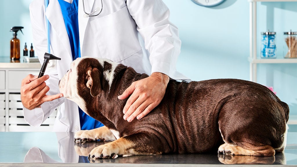
When Rudette Plant saw the pink pulpy mass forming on her English Bulldog Lola’s eyelid she didn’t panic. A longtime volunteer for various animal rescue groups, Plant had seen the defect before—it was cherry eye in dogs.
“I imagine it would be very frightening if you didn’t know what it was,” says Plant, a Garden Grove, California resident, in regard to the dime-sized, fleshy protrusion.
When Deborah Turner, an animal rescuer and advocate with Friends of Long Beach Animals of Long Beach, California, rescued a cute, little brindle Boston Terrier, she knew the defect had to be fixed or the puppy would never find a home, so she paid for a procedure with her own money.
For some particular breeds such as the Boston Terrier, cherry eye is common. Find out what cherry eye in dogs is, the causes of it and how to treat it.
What Is Cherry Eye in Dogs?
The ailment called cherry eye is a disorder of the nictitating membrane, or third eyelid. The third eyelid is the filmy part for the eye in the lower corner nearest the nose. Cherry eye in dogs occur when the gland connected to the third eyelid becomes detached and slips to protrude from the eye, presenting itself as a red pulpy mass. Depending on the severity, the protrusion of the gland can be small or as large as the tip of a thumb.
Although unsightly, cherry eye is a condition that actually looks worse than it is—as long as it is treated.
Veterinary ophthalmologists say it is a rather normal eye problem, usually occurring in younger dogs and easily corrected with surgery.
“It’s one of the much more common eye diseases in young dogs,” says Dr. D.J. Haeussler, a veterinary ophthalmologist with the Animal Eye Institute of Cincinnati, Ohio.
How Is Cherry Eye in Dogs Treated?
Unfortunately, prescription eye care for pets isn’t enough to treat cherry eye. Most often, the vet-recommended route to take is surgery, which is required to correct cherry eye.
“It absolutely needs to be addressed,” Dr. Haeussler says. “It’s not an emergency, but it should be treated surgically, particularly if it is out for a week or more.”
Dr. Nancy Bromberg, a veterinary ophthalmologist with VCA SouthPaws in Fairfax, Virginia, estimates that about 90 percent of the cases she sees require surgery at some point.
Occasionally, the protrusion will revert on its own, and, sometimes, with massage and topical applications, the protrusion will pop back into the eye. In those cases, no additional treatment is needed. But, those cases are rare.
In Lola’s case, massages worked for a couple of years, but eventually the dog eye care treatments stopped working and surgery became necessary.
“I have been able to manually reposition (the gland),” Dr. Bromberg says. “It depends how long it has been out and how swollen it is.”
Plant says she had known pet owners who didn’t treat the ailment because of the cost, which in her case was about $450 per eye.
Why Is Surgery Needed?
“The gland at the base of the eye is to produce tears,” Dr. Haeussler says, adding that it is very important to the ocular health of the dog.
The tear production supplies oxygen and nutrients. Dr. Bromberg likens it to a windshield wiper for the eye. “Without tear film, dogs can develop dry eye,” Dr. Haeussler says.
Dry eye, or keratoconjunctivitis sicca, can be an irritant and painful for the dog. In worst cases, it can lead to blurred vision or corneal scarring and blindness if the dog continually digs at the eye with its paws to ease the irritation.
“If it stays out for a long time, it becomes a vicious cycle,” says Dr. Bromberg. Surgery can immediately rectify the issue, which lessens the dog’s risk of developing dry eye.
What Is the Surgery Like?
The procedure is considered pretty straightforward with a short recovery time and few lingering side effects. There are a number of methods to reattach the gland, and opinions sometimes vary about the most effective and longest-lasting.
Dr. Haeussler says his preferred method it to “surgically prepare a pocket,” and suture the gland. He uses the “Morgan pocket,” while Dr. Bromberg says she uses a modified procedure.
Surgical reattachment is not always permanently successful, either.
“Unfortunately, it’s not 100 percent, so I warn patients,” Dr. Bromberg says, explaining that sometimes the repair breaks down over time.
“I try bringing connective tissue over the gland—that seems to be more effective,” she says. “A lot of times it depends on how large the gland is. I don’t think any two cherry eyes are exactly alike.”
Although it was once common to simply remove the gland, most ophthalmologists now repair the eye surgically. The third eyelid is small and mostly a vestigial, or no longer useful, portion of the eye. However, the tear-producing gland is vital.
“Before the importance of the gland was known, they would just clamp it and cut it out,” Dr. Bromberg says.
Many dogs require surgery to both eyes, although Dr. Bromberg said it was less than 50 percent in the cases she’s seen.
What Causes Cherry Eye?
It is unknown what is responsible for the disorder. Although not hereditary, cherry eye tends to be “breed-related,” according to Dr. Bromberg and is most common to Bulldogs and Shih-tzus.
Eventually, the gene responsible for the disorder is likely to be found, according to Dr. Bromberg.
Lola would eventually require surgery in both eyes to repair the ailments, but recovered quickly and suffered no ill effects afterward. The Boston Terrier found a home after surgery and has been symptom-free ever since.
By: Greg Mullen
Share:


